Beranda
/ Back Muscles Anatomy - Back Of Neck Anatomy / Muscles Of The Neck Laminated ... / The extrinsic back muscles are located in the back, but act to produce movements of the shoulder and assist respiration.
Back Muscles Anatomy - Back Of Neck Anatomy / Muscles Of The Neck Laminated ... / The extrinsic back muscles are located in the back, but act to produce movements of the shoulder and assist respiration.
Insurance Gas/Electricity Loans Mortgage Attorney Lawyer Donate Conference Call Degree Credit Treatment Software Classes Recovery Trading Rehab Hosting Transfer Cord Blood Claim compensation mesothelioma mesothelioma attorney Houston car accident lawyer moreno valley can you sue a doctor for wrong diagnosis doctorate in security top online doctoral programs in business educational leadership doctoral programs online car accident doctor atlanta car accident doctor atlanta accident attorney rancho Cucamonga truck accident attorney san Antonio ONLINE BUSINESS DEGREE PROGRAMS ACCREDITED online accredited psychology degree masters degree in human resources online public administration masters degree online bitcoin merchant account bitcoin merchant services compare car insurance auto insurance troy mi seo explanation digital marketing degree floridaseo company fitness showrooms stamfordct how to work more efficiently seowordpress tips meaning of seo what is an seo what does an seo do what seo stands for best seotips google seo advice seo steps, The secure cloud-based platform for smart service delivery. Safelink is used by legal, professional and financial services to protect sensitive information, accelerate business processes and increase productivity. Use Safelink to collaborate securely with clients, colleagues and external parties. Safelink has a menu of workspace types with advanced features for dispute resolution, running deals and customised client portal creation. All data is encrypted (at rest and in transit and you retain your own encryption keys. Our titan security framework ensures your data is secure and you even have the option to choose your own data location from Channel Islands, London (UK), Dublin (EU), Australia.
Back Muscles Anatomy - Back Of Neck Anatomy / Muscles Of The Neck Laminated ... / The extrinsic back muscles are located in the back, but act to produce movements of the shoulder and assist respiration.. The deep muscles develop in the back called intrinsic muscles. Understanding lower back anatomy is key to understanding the root of lower back and hip pain. All about the back muscles the back anatomy includes the latissimus dorsi, trapezius, erector spinae, rhomboid, and the teres major. The human spine is composed of 4 sections of vertebrae. The muscles of the back muscles make up a large part of the anatomy (structure) of the back.
They provide movements of the spine , stability to the trunk, as well as the coordination between the movements of the limbs and trunk. Anatomy of back muscles your back consists of three distinct layers of muscles, namely the superficial layer, the intermediate layer, and the deep layer. These are the muscles that are farther from the surface, closer to the internal organs and the spine. Understanding lower back anatomy is key to understanding the root of lower back and hip pain. These muscles give height and breadth to back development.
Back Muscle Anatomy Model - Human Anatomy from chandlerphysicaltherapy.net The human spine is composed of 4 sections of vertebrae. The muscles of the back can be arranged into 3 categories based on their location: Together these muscles form a column, known as the erector spinae. The muscles of the lower back help stabilize, rotate, flex, and extend the spinal column, which is a bony tower of 24 vertebrae that gives the body structure and houses the spinal cord. Muscles of the lumbar spine. To control the posture of the entire body. This curve, called lordosis, helps to: Superficial back muscles, intermediate back muscles and intrinsic back muscles.the intrinsic muscles are named as such because their embryological development begins in the back, oppose to the superficial and intermediate back muscles which develop elsewhere and are therefore classed as extrinsic muscles.
The muscles of the back muscles make up a large part of the anatomy (structure) of the back.
Three types of back muscles that help the spine function are extensors, flexors and obliques. The erector spinae is situated posterolaterally to spinal column, between the vertebral spinous processes and the costal angle of the ribs. For more anatomy content please follow us and visit our website: The back consists of the spine, spinal cord, muscles, ligaments, and nerves. 1 function of the back muscles. On this page, you'll learn about each of these muscles, their locations and functional anatomy. The intrinsic back muscles are found deeper to the extrinsic muscles, separated from them by the thoracolumbar fascia. The muscles of the lower back, including the erector spinae and quadratus lumborum muscles, contract to extend and laterally bend the vertebral column. The human spine is composed of 4 sections of vertebrae. The back anatomy includes the latissimus dorsi, trapezius, erector spinae, rhomboid, and the teres significant. Human musculature bodybuilding infographic muscular system vector human anatomy back muscle anatomy bicep male muscular anatomy human body anatomy female female anatomy muscle hamstrings muscle. To control the posture of the entire body. To perform clinical clinical orthopedic manual therapy to the lumbar spine.
The anatomy of the back muscles: Back muscles, functions and exercises: Leaning back to straight vertical and all points in between. These layers of back muscles help to mobilize and stabilize your trunk during your day to day activities. We hope this picture anatomy of back muscles diagram can help you study and research.
Back Muscle Anatomy Model - Human Anatomy from chandlerphysicaltherapy.net The intrinsic back muscles are found deeper to the extrinsic muscles, separated from them by the thoracolumbar fascia. The extrinsic back muscles are located in the back, but act to produce movements of the shoulder and assist respiration. Leaning back to straight vertical and all points in between. These muscles include the large paired muscles in the lower back, called erector spinae, which help hold up the spine, and gluteal muscles. Back pain is common and might be caused by a problem with a muscle. The human spine is composed of 4 sections of vertebrae. (2017, elsevier) should be consulted. The muscles of the back muscles make up a large part of the anatomy (structure) of the back.
As a general group, they extend from the neck to the sacrum and fulfill a basic and fundamental function:
Muscle or ligament strains can occur from repeated use of the muscles, or from improperly or awkwardly lifting heavy objects. Leaning back to straight vertical and all points in between. The surface muscles of the upper back include the trapezius muscles (traps) and posterior deltoids. This curve, called lordosis, helps to: The muscles of the back can be arranged into 3 categories based on their location: The muscles of the lower back help stabilize, rotate, flex, and extend the spinal column, which is a bony tower of 24 vertebrae that gives the body structure and houses the spinal cord. The erector spinae is situated posterolaterally to spinal column, between the vertebral spinous processes and the costal angle of the ribs. The intrinsic back muscles are found deeper to the extrinsic muscles, separated from them by the thoracolumbar fascia. Anatomy of the back muscles the latissimus dorsi muscles (also known as the lats) are the largest muscles of the back. The anatomy of the back muscles: These structures work together to support the body, enable a range of movements, and send messages from the brain to the. (2017, elsevier) should be consulted. These muscles give height and breadth to back development.
These muscles give height and breadth to back development. What are the lower back muscles and their anatomy? The deep muscles develop in the back called intrinsic muscles. These layers of back muscles help to mobilize and stabilize your trunk during your day to day activities. The back consists of the spine, spinal cord, muscles, ligaments, and nerves.
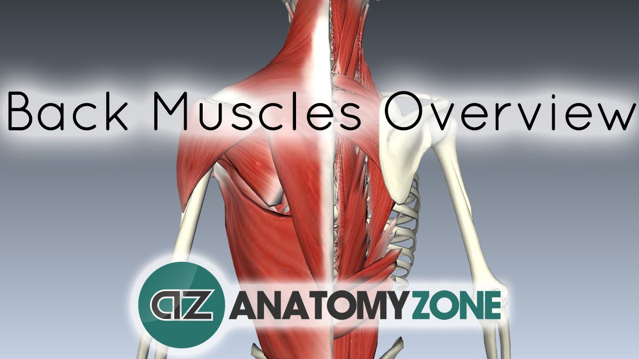
Back Muscles in a Nutshell - Anatomy Tutorial - YouTube from i.ytimg.com Human musculature bodybuilding infographic muscular system vector human anatomy back muscle anatomy bicep male muscular anatomy human body anatomy female female anatomy muscle hamstrings muscle. The back muscles are anatomically layered into superficial (extrinsic) and deep (intrinsic) muscles. On this page, youll learn about each of these muscles, their locations, and functional anatomy. They provide movements of the spine , stability to the trunk, as well as the coordination between the movements of the limbs and trunk. 4 erector spinae (back erectors). This blog post article is an overview of the muscles of the lumbar spine of the trunk. Leaning back to straight vertical and all points in between. Back pain is one of the most common kinds of pain for adults, and muscle strains are the most common type of back pain.
Anatomy of the back muscles the latissimus dorsi muscles (also known as the lats) are the largest muscles of the back.
On this page, youll learn about each of these muscles, their locations, and functional anatomy. Understanding lower back anatomy is key to understanding the root of lower back and hip pain. We hope this picture anatomy of back muscles diagram can help you study and research. The muscles of the lower back help stabilize, rotate, flex, and extend the spinal column, which is a bony tower of 24 vertebrae that gives the body structure and houses the spinal cord. The anatomy of the back muscles: Back muscles the muscles of the back are a group of strong, paired muscles that lie on the posterior aspect of the trunk. The muscles of the back muscles make up a large part of the anatomy (structure) of the back. To perform clinical clinical orthopedic manual therapy to the lumbar spine. Your lower back (lumbar spine) is the anatomic region between your lowest rib and the upper part of the buttock. To control the posture of the entire body. (2017, elsevier) should be consulted. The surface muscles of the upper back include the trapezius muscles (traps) and posterior deltoids. The muscles, bones, ligaments, and tendons in the back can all be injured and cause back pain.